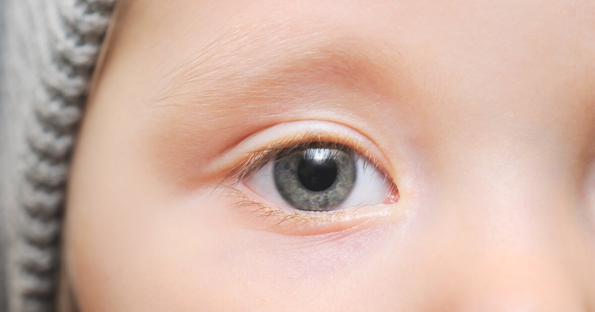
Researchers have taken photographs of children’s retinas and screened them using a deep learning AI algorithm to diagnose autism with 100% accuracy. The findings support using AI as an objective screening tool for early diagnosis, especially when access to a specialist child psychiatrist is limited.
AI-screened eye pics diagnose childhood autism with 100% accuracy::undefined
There was no notable decrease in the mean AUROC, even when 95% of the least important areas of the image – those not including the optic disc – were removed.
So we know that it relates to the optic disc.
Edit: Repeated in the conclusions of the study itself:
Our findings suggest that the optic disc area is crucial for differentiating between individuals with ASD and TD.
Edit 2: Which is given more background as to what may be going on and being picked up by the model:
Considering that a positive correlation exists between retinal nerve fiber layer (RNFL) thickness and the optic disc area,32,33 previous studies that observed reduced RNFL thickness in ASD compared with TD14-16 support the notable role of the optic disc area in screening for ASD. Given that the retina can reflect structural brain alterations as they are embryonically and anatomically connected,12 this could be corroborated by evidence that brain abnormalities associated with visual pathways are observed in ASD. First, reduced cortical thickness of the occipital lobe was identified in ASD when adjusted for sex and intelligence quotient.34 Second, ASD was associated with slower development of fractional anisotropy in the sagittal stratum where the optic radiation passes through.35 Interestingly, structural and functional abnormalities of the visual cortex and retina have been observed in mice that carry mutations in ASD-associated genes, including Fmr1, En2, and BTBR,36-38 supporting the idea that retinal alterations in ASD have their origins at a low level.

Hold the fuck up. What exactly is the marker?
A big problem with this type of ai is they are a black box.
We don’t know what they are identifying. We give it input and it gives output. What exactly is going on internally is a mystery.
Not so much of a mystery:
So we know that it relates to the optic disc.
Edit: Repeated in the conclusions of the study itself:
Edit 2: Which is given more background as to what may be going on and being picked up by the model: